Rectopen: Physiology

Download Free Android Apps

Ads 728x90

Showing posts with label Physiology. Show all posts
Digestive System
November 18, 2022
Digestive System_পরিপাকতন্ত্র
The digestive system is made up of the gastrointestinal tract—also called the GI tract or digestive tract—and the liver, pancreas, and gallbladder.

The human gastrointestinal tract, also called the alimentary canal, is around 30 feet (9 meters) long in adults.

It consists of the following:

the mouth, or oral cavity
the esophagus
the stomach
the small intestine
the large intestine, or colon
the rectum

In addition, the following organs support digestion, for example, by chewing or adding enzymes and other secretions that enable the body to absorb nutrients:

the teeth
the tongue
the salivary glands
the liver
the pancreas
the gallbladder

Together, these organs provide mechanical processing, the secretion of enzymes and bile to help break down compounds, and the excretion of waste.

Some terms to describe these functions include:

ingestion, which includes chewing and swallowing
the secretion of substances that enable food and nutrients to move through the body effectively.

propulsion, where muscles move the contents of the canal forward
digestion, the breakdown of food, for example, through chewing and secretions.

the absorption of nutrients, which mainly occurs in the small intestine
defecation and urination, the removal of waste products through the rectum and the bladder, respectively.

gastrointestinal tract
Gastrointestinal tract

পরিপাকতন্ত্রের সংজ্ঞা দাও?
মুখ থেকে মলদ্বার পর্যন্ত খাদ্যনালী এবং সংশ্লিষ্ট অঙ্গ সমবায়ে মানব পরিপাকতন্ত্র গঠিত যার মূল কাজ খাদ্য পরিপাক করা। একে পাচনতন্ত্র (digestive system) বা পৌষ্টিকতন্ত্রও বলা হয়ে থাকে। অথবা, যে তন্ত্রের মাধ্যমে পরিপাক ক্রিয়া সম্পন্ন হয় তাই পরিপাকতন্ত্র বা পৌষ্টিকতন্ত্র।
খাদ্য পাচন বা পরিপাক বা হজম একটি শারীরিক প্রক্রিয়া যার মাধ্যমে খাদ্যকে প্রথমে ছোট ছোট টুকরো করে তার পর পর্যায়ক্রমে বিভিন্ন এনজাইম Enzyme দ্বারা বিগলিত করে দেহে আত্তীকরণের বা হজমের উপযোগী করা হয়। পাচন প্রক্রিয়ায় নিযুক্ত অঙ্গতন্ত্রকে পাচনতন্ত্র বলে।

Digestive system diagram and functions

1. Oral Cavity
    -Nasal Cavity
    -Palate
    -Oral Cavity
    -Lips
    -Tongue
    -Pharynx
    -Epiglottis
    -Larynx opening into pharynx
    -Larynx
    -Esophagus

2. Tongue
Papillae: 
    -Filiform
    -Fungiform
    -Circumvallate
    -Foliate
Taste buds:
    -Sweet
    -Salty
    -Bitter
    -Sour
    -Umami (savory)

Your tongue is covered in tiny papillae (bumps) and thousands of taste buds.

3. Salivary Gland
4. Teeth
5. Oesophagus
7. Stomach
8. Small Intestine
9. Large Intestine
10. Caecum
11. Appendix
12. Colon
13. Rectum
14. Anal Canal
15. Liver
16. Gall Bladder
17. Billiary Apparatus/ Bile Duct
18. Hepatobiliary System
19. Pancreas
20. Peritoneum


পাকস্থলীর অংশগুলি কি কি?
পাকস্থলীর ওপরের অংশে আছে একটি নল যার নাম খাদ্যনালী বা ইসোফেগাস। যখন তুমি চিবোনো খাবার গিলে ফেলো তখন এই নালিকাটির মুখ খুলে যায় যাতে খাদ্য পাকস্থলীতে পৌঁছোতে পারে। পাকস্থলী অনেক পেশী দিয়ে তৈরি। এই পেশীগুলি খাদ্যকে ছোট ছোট টুকরো করে ভেঙে ফেলতে সাহায্য করে। পেশীগুলি এমন তরঙ্গ সৃষ্টি করে, যাতে খাদ্য পাচনতন্ত্রের আরও নিচের দিকে নেমে যায়। একে বলা হয় পেরিস্টলসিস। এই প্রক্রিয়া খাদ্যনালী থেকে শুরু করে বৃহদন্ত্র পর্যন্ত সমস্ত পথেই ঘটে।

পাকস্থলীর সঙ্গে সারি দিয়ে থাকে কিছু গ্রন্থি। এই বিশেষ গ্রন্থিগুলি কিছু তরল পদার্থ তৈরি করে যেগুলি পাকস্থলীতে এসে পড়ে। এই তরলকে পাচক রস বলা হয়। এই পাচক রসে অম্ল বা অ্যাসিড থাকে এবং সেটি খাবার ভাঙতে বা হজম করতে সাহায্য করে।

পাকস্থলীর নিচের অংশকে বলা হয় পাইলোরিক অঞ্চল। এই অঞ্চলের নিচের দিকে একটি প্রস্থান দ্বার রয়েছে। এই ছিদ্র দিয়ে খাদ্যাংশ ক্ষুদ্রান্ত্রের মধ্যে প্রবেশ করে।

পাকস্থলীর (এবং সাধারণত পাচনতন্ত্রের) সবচেয়ে গুরুত্বপূর্ণ অংশগুলির মধ্যে একটি হল ব্যাকটেরিয়া প্রাণী যা এইখানেই বাস করে। নির্দিষ্ট পরিবেশের মধ্যে একদম আলাদা ধরণের এই ব্যাকটেরিয়া পাচনতন্ত্রের গুরুত্বপূর্ণ অংশ হিসেবে উৎসেচক তৈরি করে। সেগুলি কিছু খাদ্যের পাচনের জন্য প্রয়োজন হয়। এছাড়াও আমরা যা আহার করি তার সর্বাধিক পুষ্টি অংশ যাতে নিষ্কাশিত করা যায়, তাতেও সহায়তা করে। এই ব্যাকটেরিয়াগুলির সঙ্গে আমাদের একটি মিথোজীবী (পারস্পরিক উপকারী) সম্পর্ক থাকে যা শুরু হয় জন্ম থেকেই, মায়ের কাছ থেকে শিশু যে দুধ পায় তার মাধ্যমে এগুলি শিশুর শরীরে চলে আসে এবং বয়সের সঙ্গে সঙ্গে তার আধুনিকীকরণও হতে থাকে।

পাকস্থলীর কাজ কি?
গ্রহণ করা খাদ্য পাকস্থলীতে এসে সঞ্চিত হয় এবং সেখানে এর পাচন ক্রিয়া শুরু হয়। পাকস্থলী তার পেশীগুলির সংকোচন ও প্রসারণ করে খাদ্যকে ছোট ছোট টুকরো টুকরো করে এই কাজ করে। এছাড়াও খাদ্যকে ভেঙ্গে দিতে পাকস্থলী পাচক উৎসেচক এবং অম্লরস ব্যবহার করে।

পাকস্থলী কিভাবে শরীরের অন্যান্য অংশের সাথে যোগাযোগ রাখে ?
তুমি কিছু চিন্তা করার আগেই পাকস্থলী তার নিজের কাজ করে ফেলে। এই কাজটি স্নায়ুতন্ত্র দ্বারা নিয়ন্ত্রিত হয়। পাকস্থলী খালি হয়ে গেলে স্নায়ু তোমার মধ্যে ক্ষিদের অনুভূতি আনতে সাহায্য করে। তারা তোমার পাকস্থলীকেও সংকেত পাঠায় যে কখন পাচক ক্রিয়া শুরু করার জন্য সে আরও রাসায়নিক তৈরি করা শুরু করবে।

পাকস্থলীর ভিতরের আস্তরণে রক্তবাহী নালী থাকে। এই নালীর মাধ্যমে রক্ত পাকস্থলীতে অক্সিজেন সরবরাহ করে যাতে এটি নিজের কাজ করতে পারে।

পাকস্থলীর পেশীগুলি খাদ্য স্থানান্তর করতে সহায়তা করে। পাকস্থলী তার কাজ সম্পন্ন করার পর, খাদ্য ক্ষুদ্রান্ত্রে চলে যায়।

কিভাবে তুমি তোমার পাকস্থলীকে সুস্থ রাখতে পারো ?
ভাল পাচন ক্রিয়া শুরু হয় চিবোনোর মাধ্যমে। খাবার ভালোভাবে চিবোনো এবং ধীরে ধীরে খাওয়া তোমার পাকস্থলীর জন্য ভাল।

ক্ষিদে পেলে তবে তুমি খাও। খুব বেশি বা খুব কম খাবার খাবে না। খুব বেশি চর্বি বা চিনি নেই এমন খাবার খাওয়ার চেষ্টা করো। তোমার খাদ্যতালিকায় শাকসবজি এবং ফল অন্তর্ভুক্ত করো। যদি কোন খাদ্য থেকে প্রায়শই তোমার পেটে ব্যথা করে বা খারাপ লাগে এবং হজমের সমস্যা হয়, তাহলে সেই খাবার এড়িয়ে চলো।

মুখগহ্বরে খাদ্য পরিপাক
আমরা সামান্য খাবার মুখে দেয়ার পর থেকেই মূলত এই তন্ত্রের কাজ শুরু হয়ে যায়। মুখের ভেতরের খাবারের অংশকে দাঁতের সাহায্যে টুকরো করা, লালাগ্রন্থি থেকে রস ক্ষরণ ও মিশ্রিত হওয়া, খাদ্যকে খাদ্যমন্ডে পরিণত করার মত পরিপাকের কাজগুলো মুখের ভেতরেই সম্পন্ন করে থাকে পরিপাকতন্ত্র।

পাকস্থলীতে খাদ্য পরিপাক
মুখ থেকে গলঃধকৃত খাবারগুলো পাকস্থলীতে এসে প্রায় ২-৬ ঘন্টা জুড়ে অবস্থান করে। এসময় শুরু হয় পাকস্থলীতে খাদ্য পরিপাকের কাজ। পাকস্থলী থেকে হাইড্রোক্লোরিক এসিড (HCl) ক্ষরণ হয়, যা খাদ্যের সকল ব্যাকটেরিয়া ধ্বংস করে থাকে। পাকস্থলীতে সকল খাদ্য এক ধরণের পেস্টস্বরূপ আকার ধারণ করে। খাদ্যের এই অবস্থা কাইম বা মন্ড নামে পরিচিত। পাকস্থলীতে রাসায়নিক পরিপাকের অংশ হিসেবে পরিপাকতন্ত্র কতৃক এক ধরণের রস নিঃসরণ করা হয়, যার নাম পাকরস। পাকরসে মিশ্রিত খাদ্য পরবর্তীতে দেহের বিভিন্ন অংশ যেমন যকৃত, ডিওডেনাম, অগ্ন্যাশয় এ পৌঁছায়। এভাবেই পরিপাকতন্ত্র বা পৌষ্টিকতন্ত্র খাদ্য পরিপাকে সহায়তা করে।

Digestive_system_diagram

পরিপাক তন্ত্রঃ
মানুষ সাধারনত যা খাদ্য গ্রহন করে যে সকল অঙ্গ একত্রে পরিপাক প্রক্রিয়াকরণে সাহায্য করে তাকে পরিপাকতন্ত্র বলে।আমরা মুখের মাধ্যমে যে খাদ্য গ্রহন করি তা অন্ত্রের মাধ্যমে পাকস্থলিতে পৌছানোর পর তা পাকস্থলি ভেঙ্গে হজমে সাহায্য করে এবং উচ্ছিষ্ট পায়ু পথের মাধ্যমে শরীর থেকে বেরিয়ে আসে। পরিপাকতন্ত্র অনেক অঙ্গের সমন্বয়ে গঠিত। 
নিম্নে কিছু পরিপাকতন্ত্রের অংশসমূহ নাম ও তাদের কার্যকর্ম দেওয়া হলোঃ
অন্ননালি - পেশল সংকোচনের মাধ্যমে খাদ্যদ্রব্য এ নালীপথে পাকস্থলিতে প্রবেশ করে।
পাকস্থলি - বিভিন্ন এনজাইম এবং এসিডের মাধ্যমে জটিল খাদ্য ভাঙ্গে।
যকৃত - পাচক" রস উৎপন্ন করে যা ফ্যাটকে ভাঙ্গে
পিত্তথলি - পাচক রস সংরক্ষন করে এবং প্রয়োজনে নিঃসরণ করে।
অগ্ন্যাশয় - একধরনের রাসায়নিক পদার্থ নিঃসরন করে যা খাদ্যকে ভাঙ্গে।
ক্ষুদ্রান্ত্র - খাদ্য শোষণ করে শরীরের জন্য।
বৃহাদান্ত্র - পানি ও লবন শোষণ করে।
মলাশয় - উচ্ছিষ্ট সরবরাহ করে।
অ্যাপেনডিক্স - চিহ্নিত অঙ্গ (এর কোনে কাজ মানব শরীরে নেই, এটি একটি অতিরিক্ত অঙ্গ)।


পাকস্থলীর আকৃতি কেমন?
পাকস্থলীর আকৃতি অনেকটা ইংরেজি 'জে' "J" অক্ষরের মতো।
পাকস্থলী অবস্থান ?
পাকস্থলীর প্রকৃত অবস্থান পাঁজরের নিচের অংশের পেছনের দিকে। পাকস্থলীকে বলা যায় একটি থলি, এটি তৈরি হয় অনেকগুলি পেশী দিয়ে। এটি আকারে হাতের মুষ্টির সমান কিন্তু খাবার বেশি থাকলে পাকস্থলী প্রসারিত হয়ে খাবারের জন্য জায়গা করে দিতে পারে। এটিতে একটি প্রবেশ entry এবং প্রস্থান exit দ্বারও রয়েছে।
পরিপাকতন্ত্রের উদ্দেশ্য কি: হজমের উদ্দেশ্য হচ্ছে
শরীরের বিভিন্ন কার্যক্রমের জন্য শক্তি যোগানো,
দেহের বৃদ্ধি করা ,
সার্বক্ষণিক ক্ষয়ের ক্ষতিপূরণ করা,
প্রজনন ও দুধ উৎপাদনের সহায়তা করা।
পরিপাকতন্ত্রের কাজ :
খাদ্য গ্রহণ,
খাদ্য পরিপাক,
বিভিন্ন পাচক রস নিঃসরণ,
পানি, ভিটামিন, খনিজ লবণ এবং পরিপাক কৃত খাদ্যাংশ শোষণ,
অপ্রয়োজনীয় খাদ্যাংশ মল হিসাবে ত্যাগ,
শরীরের পানি, গ্লুকোজ, এসিড ক্ষারের ভারসাম্য রক্ষা করা,
পরিপাকতন্ত্রকে কেন দ্বিতীয় মস্তিষ্ক বলা হয়?
আমাদের অন্ত্র লাখো নিউরনের সঙ্গে সংযুক্ত, যে কারণে অন্ত্রকে মানবদেহের "দ্বিতীয় মস্তিষ্ক" হিসেবে ডাকা হয়।
খাদ্য পরিপাক হয়? উঃ ডিওডেনাম
পৌষ্টিক নালির সাথে কয়টি গ্রন্থি আছে? উঃ ৩টি
মানুষের লালাগ্রন্থি কয় জোড়া? উঃ ৩ জোড়া
মানুষের স্থায়ী দাত কয়টি? উঃ ৩২টি
মানবদেহের পরিপাকের সাথে জড়িত অংশসমূহকে কয়টি ভাগে ভাগ করা হয় ? 
দুটি ভাগে তা হলো― ১. পৌষ্টিকনালী ২. পৌষ্টিক গ্রন্থি।
পৌষ্টিকনালী―  মুখ বিবর, গলবিল, অন্ননাল, পাকস্থলী, ক্ষুদ্রান্ত্র, বৃহদান্ত্র, পৌষ্টিক গ্রন্থি
মুখবিবরের লালা গ্রন্থি―  যকৃৎ , অগ্ন্যাশয় 
পাকস্থলীতে কিভাবে খাদ্য হজম হয়?
পাকস্থলীতে খাবার পরিপাকের জন্য হাইড্রোক্লোরিক এসিড ও পেপসিন নামক এনজাইম নিঃসৃত হয়। এসব এনজাইম আমিষ জাতীয় খাদ্য কে ভেঙ্গে পলিপেপটাইড এ পরিণত হয়। কিছু শর্করা এবং স্নেহ জাতীয় খাদ্য সাধারণত পাকস্থলীতে পরিপাক হয় না।
স্টমাক এর কাজ কি?
পাকস্থলী (ইংরেজি: Stomach) মানব দেহে পরিপাকতন্ত্রের একটি গুরুত্বপূর্ণ অংশ যা অন্ননালী ও ক্ষুদ্রান্ত্রের মধ্যে অবস্থিত। এটি উদর গহবরের বাম পাশে উপর দিকে থাকে। খাদ্য পরিপাক প্রক্রিয়া প্রধানত পাকস্থলীতে শুরু হয়। বিশেষ করে আমিষ জাতীয় খাদ্যের পরিপাকে পাকস্থলীর ভূমিকা প্রধান।
লালার এনজাইম এর নাম কি?
লালা হল একটি শারীরিয় তরল বস্তু যা প্রানীর মুখের লালা গ্রন্থি হতে উৎপন্ন হয়। মানব লালার ৯৯.৫% পানি সহ ইলেক্ট্রোলাইট, শ্লেষ্মা, শ্বেত লোহিত কনিকা, এপিথেলিয়াল কোষ (যা থেকে ডিএনএ বের করা যাবে), এনজাইম (যেমন এ্যামিলেস এবং লাইপেজ), এন্টিমাইক্রোবিয়াল এজেন্ট নি:সৃরক যেমন ইমিউনোগ্লোবিউলিন এ এবং লাইসোজাইম।
সব ধরনের খাদ্যের চূড়ান্ত পরিপাক কোথায় ঘটে?
ক্ষুদ্রান্ত্রের আকার বড় হওয়ার কারণ এর তিনটি অংশ রয়েছে এবং সব ধরনের খাদ্যের চূড়ান্ত পরিপাকই ক্ষুদ্রান্ত্রে সংঘটিত হয়।
মিউকোসা কি?
ক্ষুদ্রান্ত্র (ইংরেজি: Small intestine) মানবদেহের খাদ্য পরিপাকতন্ত্রের একটি অংশবিশেষ যা ওপরে পাকস্থলী এবং নিম্নে বৃহদন্ত্র অবস্থিত। মানবদেহের পরিপাক তন্ত্রের শুরু মুখগহ্বরে, এরপর ধারাবাহিকভাবে গ্রাসনালী, পাকস্থলী, ক্ষুদ্রান্ত্র, বৃহদন্ত্র ও মলাশয় অবস্থিত।
মানুষের পাকস্থলীর রসের pH কত?
পাকস্থলীর পাচক রসে pH এর মান 1.4 হলে ওই রসে হাইড্রোজেন আয়নের ঘনমাত্রা কত?
পানি শোষণের প্রধান স্থান কোনটি?
বৃহদন্ত্র, বৃহৎ অন্ত্র নামেও পরিচিত, যা গ্যাস্ট্রোইন্টেস্টাইনাল নালীর এবং মেরুদন্ডীদের পরিপাকতন্ত্রের শেষ অংশ। পানি এখানে শোষিত হয় এবং অবশিষ্ট বর্জ্য পদার্থ মল হিসেবে সংরক্ষণ করে মলত্যাগ এর পূর্ব পর্যন্ত।
পাকস্থলীর ক্যান্সারের লক্ষণ কি?
প্রাথমিক লক্ষণগুলো হলো বুকজ্বালা, পেটের উপরের অংশে ব্যথা, বমি ও ক্ষুধামন্দা পরবর্তী লক্ষণগুলোর মধ্যে রয়েছে ওজন কমে যাওয়া, জন্ডিস, বমি ডিসফ্যাজিয়া বা খাবার গিলতে কষ্ট হওয়া, পায়খানার সাথে কালো রক্ত যাওয়া(melena) ইত্যাদি।
ভিলাই কি এবং এর কাজ কি?
পরিপাকতন্ত্রের ক্ষুদ্রান্ত্রের মিউকোসা হতে আঙুলের ন্যায় কতগুলো অভিক্ষেপ সৃষ্টি হয় এদের ভিলাই বলে। ভিলাই এর কাজ হচ্ছে শোষণ অঞ্চল বৃদ্ধি করা।
ভিলাই-এর অবস্থান কোথায়?
ক্ষুদ্রান্ত্রের ইলিয়াম অঞ্চলে
ব্রুনার্স গ্রন্থি কোথায় পাওয়া যায়?
ব্রুনারের গ্রন্থি বা ডিওডেনামের গ্রন্থি (ইংরেজি: Brunner's gland/Duodenal gland) হল মানব পরিপাকতন্ত্রের ক্ষুদ্রান্ত্রের ডিওডেনামে অবস্থিত গ্রন্থি।
বেশি ভাজা ও তেলযুক্ত খাবার পাকস্থলীর জন্য ক্ষতিকর কেন?
তেলে ভাজা যেকোন খাবারই পাকস্থলীর উপর বাড়তি চাপ তৈরি করে এবং অ্যাসিড রিফ্ল্যাক্স ও বুক জ্বালাপোড়ার সমস্যা তৈরি করে। এছাড়া প্রক্রিয়াজাত খাবারের মতোই তেলে ভাজা খাবার তলপেটের চর্বি বৃদ্ধি করে এবং খাদ্য পরিপাক হতে লম্বা সময়ের প্রয়োজন হয়।
লালাগ্রন্থি থেকে নিঃসৃত উৎসেচকের নাম কি?
অ্যামাইলেজ (Amylase) 
মাতৃদুগ্ধের pH এর মান কত?
প্রধান দরকারি হচ্ছে শক্তি(লিপিড, ল্যাক্টোজ ও আমিষ), আমিষ (প্রয়োজনীয় এ্যামিনো এসিড এবং এ্যামিনো দল বা গ্রুপ) এর দ্বারা অনৈত্যাবশ্যকীয় এ্যামিনো এসিডের জৈব সংশ্লেষণ সরবরাহ, প্রয়োজনীয় ফ্যাটি এসিড, ভিটামিন, অজৈব উপাদান এবং পানি। পি. এইচ দুধের পি. এইচ(pH) সীমা ৬.৪ থেকে ৬.৮ পর্যন্ত এবং সময়ের সাথে পরিবর্তিত হয়।
সিকামের অবস্থান কোথায়?
পাচনতন্ত্রে বৃহদন্ত্রের প্রথম ভাগ। ক্ষুদ্রান্ত্র আসলে বৃহদন্ত্রের একদম গোড়াতে প্রবেশ করে না। ক্ষুদ্রান্ত্রের পরেই খাদ্য বৃহদন্ত্রের অ্যাসেন্ডিং কোলন অংশে সম্মুখচালিত হয়। কিন্তু ক্ষুদ্রান্ত্র আর বৃহদন্ত্রের সংযোগস্থল থেকে বিপরীত গামী অন্ধ-গলি (ব্লাইন্ড পাউচ) হল সিকাম।
শর্করা ভেঙে কি তৈরি হয়?
শর্করা পরিপাক হল এমন একটি শারীরবৃত্তীয় প্রক্রিয়া যার মাধ্যমে পৌষ্টিক নালীর বিভিন্ন অংশ থেকে নিঃসৃত পাকরসে অবস্থিত শর্করা বিশ্লেষণকারী উৎসেচকের সাহায্যে বিভিন্ন শর্করা বিশ্লিষ্ট হয়ে একক শর্করা উৎপন্ন হয়। শর্করা জাতীয় খাদ্যের পরিপাক মুখগহ্বরে শুরু হয় এবং ক্ষুদ্রান্ত্রে শেষ হয়।
হরমোন কত প্রকার ও কি কি?
রাসায়নিক গঠন অনুযায়ী তিনটি প্রধান ধরনের হরমোন আছে। ১, প্রোটিন হরমোন (বা পলিপেপটাইড হরমোন) অ্যামিনো অ্যাসিডের চেইন দিয়ে তৈরি। একটি উদাহরণ হল ADH (অ্যান্টিডিউরেটিক হরমোন) যা রক্তচাপ হ্রাস করে। ৩, অ্যামাইন হরমোন অ্যামিনো অ্যাসিড থেকে উদ্ভূত হয়।
ভাজা জিনিস খেলে কি হয়?
ভাজা খাবারের তেলে থাকে ট্রান্স ফ্যাট শরীরে এলডিএলের মাত্রা বাড়ায়। এলডিএল বা 'লো-ডেনসিটি লিপোপ্রোটিন' যা খারাপ কোলেস্টেরল হিসেবে চিহ্নিত। যে কারণে ধমনীতে বাধা তৈরি করে হৃদরোগের ঝুঁকি বাড়ে। চিকিৎসা বিজ্ঞানে একটি বিষয় প্রমাণিত যে, অতিরিক্ত ভাজা খাবার খেলে রক্তচাপও বৃদ্ধি পায়, যা হৃদরোগ হওয়ার আরেক কারণ।
লালারসে কত ভাগ পানি থাকে?
এতে থাকে শতকরা ৯৮ ভাগ পানি ও ২ ভাগ এনজাইম বা জৈব রাসায়নিক পদার্থ। লালার এনজাইমকে বলে টায়ালিন। লালার জীবাণু-প্রতিরোধী ক্ষমতা আছে।
লালার প্রধান কাজ কী?
লালা জীবাণু ধ্বংস করে লালার জীবাণু-প্রতিরোধী ক্ষমতা আছে। মুখের ভেতরকার জীবাণুকে মেরে ফেলতে এটি গুরুত্বপূর্ণ ভূমিকা রেখে থাকে। বিভিন্ন সময় বিভিন্ন ভাবে আমাদের মুখে জীবাণু প্রবেশ করতে পারে।
লালাগ্রন্থি কোথায় অবস্থিত?
কানের সামনে অবস্থিত চোয়ালের দুই পাশের প্যারোটিড গ্রন্থি, মুখের নিচে দুই পাশে সাব ম্যান্ডিবুলার ও জিবের নিচের দুই পাশে সাব লিঙ্গুয়াল গ্রন্থিসহ মুখের ভেতরে অসংখ্য অতি ক্ষুদ্র লালাগ্রন্থি থেকে এটি তৈরি হয়। প্রতিদিন আধা থেকে দেড় লিটার লালা মুখের ভেতর লেগে থাকা খাবার ও জীবাণুকে পরিষ্কার ও জীবাণুমুক্ত করতে ব্যবহৃত হয়।


Lymphatic System
January 30, 2021
Table of Contents: 
  1. Definition-Lymph, Lymph nodes, Lymphatic system
  2. Parts of the lymphatic system
  3. Functions of lymphatic system
  4. Lymphatic disorders/ diseases
Lymphatic System
Lymphatic System


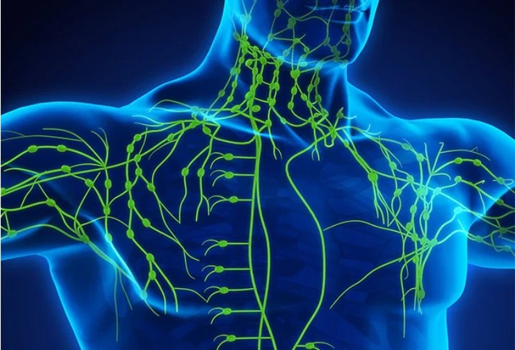
The lymphatic system is a network of tissues and organs that help rid the body of toxins, waste and other unwanted materials. The primary function of the lymphatic system is to transport lymph, a fluid containing infection-fighting white blood cells, throughout the body.


Lymphatic System
Lymphatic System

Major Organs of Lymphatic System
  1. Lymphatic vessels
  2. Lymph nodes
  3. Thymus
  4. Spleen
Major Functions of Lymphatic System
  1. Return tissue fluid to blood
  2. Carry certain absorbed food molecules
  3. Defend the body against infection
Structure of the Lymphatic System

Lymph
Lymph (from Latin, lympha meaning "water") is the fluid that flows through the lymphatic system, a system composed of lymph vessels (channels) and intervening lymph nodes whose function, like the venous system, is to return fluid from the tissues to the central circulation.
  • Lymph is a clear, colorless, water fluid derived from tissue fluid and contained within lymph vessels.
  • Lymph is similar in composition to blood except that it has a lower concentration of plasma proteins
Lymph nodes
Lymph nodes are small glands that filter lymph, the clear fluid that circulates through the lymphatic system. They become swollen in response to infection and tumors. Lymphatic fluid circulates through the lymphatic system, which is made of channels throughout your body that are similar to blood vessels.

Lymph nodes
Lymph nodes


Lymph nodes are bean-shaped glands that monitor and cleanse the lymph as it filters through them. The nodes filter out the damaged cells and cancer cells. These lymph nodes also produce and store lymphocytes and other immune system cells that attack and destroy bacteria and other harmful substances in the fluid. 

You have about 600 lymph nodes scattered throughout your body. Some exist as a single node; others are closely connected groups called chains. A few of the more familiar locations of lymph nodes are in your armpit, groin and neck. Lymph nodes are connected to others by the lymphatic vessels.

Parts of the lymphatic system


Lymph, also called lymphatic fluid, is a collection of the extra fluid that drains from cells and tissues (that is not reabsorbed into the capillaries) plus other substances. The other substances include proteins, minerals, fats, nutrients, damaged cells, cancer cells and foreign invaders (bacteria, viruses, etc.). Lymph also transports infection-fighting white blood cells (lymphocytes).

Composition of lymph fluid

Composition of lymph fluid

Lymphatic vessels:

Lymphatic vessels
Lymphatic vessels


Lymphatic vessels are the network of capillaries (micro vessels) and large network of tubes located throughout the body that transport lymph away from tissues. Lymphatic vessels collect and filter lymph (at the nodes) as it continues to move toward larger vessels called collecting ducts. These vessels operate very much like your veins do: they work under very low pressure, have a series of valves in them to keep the fluid moving in one direction.

Spleen:
This largest lymphatic organ is located on your left side under your ribs and above your stomach. The spleen filters and stores blood and produces white blood cells that fight infection or disease.
Spleen
Spleen

Thymus:
This organ is located in the upper chest beneath the breast bone. It matures a specific type of white blood cell that fights off foreign organisms.


Thymus gland
Thymus gland


Functions of Thymus
1. Primary function-production of thymic lymphocytes.
2. A major organ for proliferation of lymphocytes in body.
3. Plays key role in determining the differentiation of T cell.
4. Lymphocytes during maturation acquire new surface antigens (The Ag) & are called as T lymphocytes or T cells (thymus dependent).
5. Lymphocyte proliferation in thymus is not dependent on antigenic stimulus (unlike in peripheral lymphoid organs).
6. T lymphocytes-primarily responsible for Cell-Mediated
7. Immunity


Tonsils and adenoid:

Tonsils and adenoid
Tonsils and adenoid

These lymphoid organs trap pathogens from the food you eat and air you breathe. They are your body’s first line of defense against foreign invaders.

Bone marrow:
This is the soft, spongy tissue in the center of certain bones, such as the hip bone and breastbone. White blood cells, red blood cells, and platelets are made in the bone marrow.


Bone marrow
Bone marrow

Peyer’s patches:
These are small masses of lymphatic tissue in the mucous membrane that lines your small intestine. These lymphoid cells monitor and destroy bacteria in the intestines.

Peyer’s patches
Peyer’s patches

Appendix:
Your appendix contains lymphoid tissue that can destroy bacteria before it breaches the intestine wall during absorption. Scientists also believe the appendix plays a role in housing “good bacteria” and repopulating our gut with good bacteria after an infection has cleared.

Appendix
Appendix
Functions of lymphatic system
It maintains the balance of fluid between the blood and tissues, known as fluid homeostasis.
It facilitates absorption of fats and fat-soluble nutrients in the digestive system.

  1. Lymph carries protein and large particulate matter away from the tissue space.
  2. End products of digestion are absorbed mainly by lymph channels.
  3. Important role in redistribution of fluid in the body.
  4. Bacteria, toxins and other foreign bodies are removed from the tissues.
  5. Maintenance of structural and functional integrity of tissue.
  6. In immune response of the body.
  7. Production and maturation of lymphocytes.

It forms part of the body’s immune system and helps defend against bacteria and other intruders.

Transports and removes waste products and abnormal cells from the lymph.
  1. Transport clean fluids back to the blood
  2. Drains excess fluids from tissues
  3. Removes debris from cells of body
  4. Transports fats from digestive system
  5. It is responsible for the removal of interstitial fluid from tissues.
  6. It transports white blood cells to and from the lymph nodes into the bones.
  7. The lymph transports antigen presenting cells, such as dendrite cells to the

lymph nodes where an immune response is stimulated. Production of immune cells (such as lymphocytes, monocytes, and antibody producing cells called plasma cells).
Lymphatic disorders/diseases:

Lymphatic disorders/diseases


Immune System
January 29, 2021
  • Components of the immune system
  • Definition and classification of immunity
  • Antigen
  • Antibody
  • Antigen –antibody reaction
  • Immunodeficiency  disorders
Immune System
Immune System


Components of the Immune system

  • White blood cells
  • Antibodies
  • Complement system
  • Lymphatic system
  • Spleen
  • Bone marrow
  • Thymus.
Immune System
Immune System


White blood cells
  • White blood cells are the key players in your immune system. They are made in your bone marrow and are part of the lymphatic system. 
  • White blood cells move through blood and tissue throughout your body, looking for foreign invaders (microbes) such as bacteria, viruses, parasites and fungi. When they find them, they launch an immune attack. 
  • White blood cells include lymphocytes (such as B-cells, T-cells and natural killer cells), and many other types of immune cells. 


Antibodies
Antibodies help the body to fight microbes or the toxins (poisons) they produce. They do this by recognizing substances called antigens on the surface of the microbe, or in the chemicals they produce, which mark the microbe or toxin as being foreign. The antibodies then mark these antigens for destruction. There are many cells, proteins and chemicals involved in this attack. 

Complement system
The complement system is made up of proteins whose actions complement the work done by antibodies.

Lymphatic system
The lymphatic system is a network of delicate tubes throughout the body. The main roles of the lymphatic system are to:
  • manage the fluid levels in the body
  • react to bacteria
  • deal with cancer cells 
  • deal with cell products that otherwise would result in disease or disorders
  • absorb some of the fats in our diet from the intestine. 
The lymphatic system is made up of:
  • lymph nodes (also called lymph glands) -- which trap microbes
  • lymph vessels -- tubes that carry lymph, the colorless fluid that bathes your body's tissues and contains infection-fighting white blood cells
  • white blood cells (lymphocytes).


Special sense of Ear
December 27, 2020
The ear is the organ of hearing and balance. The parts of the ear include:

Ear
Ear

External or outer ear, consisting of:
  • Pinna or auricle. This is the outside part of the ear.
  • External auditory canal or tube. This is the tube that connects the outer ear to the inside or middle ear.
    Ear
    Ear
External auditory canal- also called external auditory meatus, or external acoustic meatus, passageway that leads from the outside of the head to the tympanic membrane, or eardrum membrane, of each ear. ... The canal is nearly 1 inch (2.5 cm) in length and is lined with skin that extends to cover the tympanic membrane.

Pinna or auricle: The medical term for the outer ear is the auricle or pinna. The outer ear is made up of cartilage and skin. There are three different parts to the outer ear; the tragus, helix and the lobule. The ear canal starts at the outer ear and ends at the ear drum.

Middle ear (tympanic cavity), consisting of:
  • Tympanic membrane (eardrum). The tympanic membrane divides the external ear from the middle ear.
  • Ossicles. Three small bones that are connected and transmit the sound waves to the inner ear. The bones are called:
  1. Malleus
  2. Incus
  3. Stapes
Tympanic membrane- also called eardrum, thin layer of tissue in the human ear that receives sound vibrations from the outer air and transmits them to the auditory ossicles, which are tiny bones in the tympanic (middle-ear) cavity.

Ossicles: The ossicles (also called auditory ossicles) are three bones in either middle ear that are among the smallest bones in the human body. They serve to transmit sounds from the air to the fluid-filled labyrinth (cochlea). The absence of the auditory ossicles would constitute a moderate-to-severe hearing loss.

Inner ear, consisting of:
  • Cochlea- This contains the nerves for hearing.
  • Vestibule- This contains receptors for balance.
  • Semicircular canals- This contains receptors for balance.
  • Eustachian tube- A canal that links the middle ear with the back of the nose. The eustachian tube helps to equalize the pressure in the middle ear. Equalized pressure is needed for the proper transfer of sound waves. The eustachian tube is lined with mucous, just like the inside of the nose and throat.
  • Vestibulocochlear nerve-

Eustachian tube, The Eustachian tube is a canal that connects the middle ear to the nasopharynx, which consists of the upper throat and the back of the nasal cavity. It controls the pressure within the middle ear, making it equal with the air pressure outside the body. Blocked Eustachian tubes can cause pain, hearing difficulties, and a feeling of fullness in the ears. Such a phenomenon is referred to as Eustachian tube dysfunction (ETD).

Vestibule- The vestibule is the central part of the bony labyrinth in the inner ear, and is situated medial to the eardrum (tympanic cavity), behind the cochlea, and in front of the three semicircular canals. The name comes from the Latin vestibulum, literally an entrance hall.

Cochlea:
  • The cochlea is a portion of the inner ear that looks like a snail shell (cochlea is Greek for snail.) 
  • The cochlea is made up of three canals wrapped around a bony axis, the modiolus. These canals are: the scala tympani , the scala vestibuli  and the scala media (or cochlear duct) .
  • The cochlea is filled with fluid. Specifically, the scala vestibuli and scala tympani contain a fluid called perilymph, which is similar in composition to cerebrospinal fluid, and the scala media contains endolymph, which more resembles intracellular fluid in terms of its ionic concentrations.
  • The cochlea contains the spiral organ of Corti, which is the receptor organ for hearing. It consists of tiny hair cells that translate the fluid vibration of sounds from its surrounding ducts into electrical impulses that are carried to the brain by sensory nerves.
  • The auditory nerve is a bundle of nerve fibres that carry information between the cochlea in the inner ear and the brain. The function of the auditory nerve is to transmit signals from the internal ear to the brain.

Semicircular canals- Semicircular canals are three tiny, fluid-filled tubes in your inner ear that help you keep your balance. When your head moves around, the liquid inside the semicircular canals sloshes around and moves the tiny hairs that line each canal.

Cochlear Damage- It is the main organ of hearing and is part of your inner ear. Cochlear Damage means that all or part of your inner ear has been hurt. Damage to the cochlea typically causes permanent hearing loss. This is called sensori neural hearing loss (SNHL).

Mechanism of hearing:
Hearing starts with the outer ear. When a sound is made outside the outer ear, the sound waves, or vibrations, travel down the external auditory canal and strike the eardrum (tympanic membrane). The eardrum vibrates. The vibrations are then passed to 3 tiny bones in the middle ear called the ossicles. The ossicles amplify the sound. They send the sound waves to the inner ear and into the fluid-filled hearing organ (cochlea).

Once the sound waves reach the inner ear, they are converted into electrical impulses. The auditory nerve sends these impulses to the brain. The brain then translates these electrical impulses as sound.
  • External ear "collects" the sound
  • Ossicles vibrate in the air-filled environment, magnifying the sound and aiming it to the oval window
  • The cochlea converts a mechanical stimulus into an electrical stimulus into an electrical one
  • The message is transported to the brain by the VIII-th nerve


Any vibrating object causes waves of compression and rarefaction and is capable of producing sound.

Sound travels faster in liquids and solids than in air (roughly 344 m per second)

When sound energy has to pass from air to liquid, most of it is reflected because of impedance by the liquid

Mechanism of hearing can be broadly classified into:
  1. Mechanical conduction of sound
  2. Transduction of mechanical energy into electrical impulses.
  3. Conduction of electrical impulses to brain.


Hormones
December 23, 2020

What is hormone ?

Hormones are chemical messengers that are secreted directly into the blood, which carries them to organs and tissues of the body to exert their functions. There are many types of hormones that act on different aspects of bodily functions and processes. Some of these include:

  1. Development and growth
  2. Metabolism of food items
  3. Sexual function and reproductive growth and health
  4. Cognitive function and mood
  5. Maintenance of body temperature and thirst

Hormones
Hormones


Where are they secreted from?

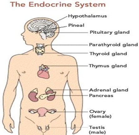
Hormones are secreted from the endocrine glands in the body. The glands are ductless, so hormones are secreted directly into the blood stream rather than by way of ducts. Some of the major endocrine glands in the body include:
  1. Pituitary gland
  2. Pineal gland
  3. Thymus
  4. Thyroid
  5. Adrenal glands
  6. Pancreas
  7. Testes
  8. Ovaries


These organs secrete hormone in microscopic amounts and it takes only very small amounts to bring about major changes in the body. Even a very slight excess of hormone secretion can lead to disease states, as can the slightest deficiency in a hormone.

List of hormone:
  • Adrenaline
  • Adrenocorticotropic hormone
  • Aldosterone
  • Androstenedione
  • Angiotensin
  • Anti-diuretic hormone
  • Anti-Müllerian hormone
  • Calcitonin
  • Cholecystokinin
  • Corticotrophin-releasing hormone
  • Cortisol
  • Dehydroepiandrosterone
  • Dihydrotestosterone
  • Erythropoietin
  • Follicle stimulating hormone
  • Gastrin
  • Ghrelin
  • Glucagon
  • Glucagon-like peptide 1
  • Glucose-dependent insulinotropic peptide
  • Gonadotrophin-releasing hormone
  • Growth hormone
  • Growth hormone-releasing hormone
  • Human chorionic gonadotrophin
  • Insulin
  • Kisspeptin
  • Leptin
  • Luteinising hormone
  • Melanocyte-stimulating hormone
  • Melatonin
  • Oestradiol
  • Oestriol
  • Oestrone
  • Oxytocin
  • Parathyroid hormone
  • Peptide YY
  • Progesterone
  • Prolactin
  • Prostaglandins
  • Relaxin
  • Somatostatin
  • Testosterone
  • Thyroid stimulating hormone
  • Thyrotropin-releasing hormone
  • Thyroxine
  • Triiodothyronine
  • Vitamin D

Endocrine system:

The hormones produced in the hypothalamus are:
  1. Corticotrophin-releasing hormone
  2. Dopamine
  3. Growth hormone-releasing hormone
  4. Somatostatin
  5. Gonadotrophin-releasing hormone
  6. Thyrotrophin-releasing hormone.


Question and Answer of Physiology
November 26, 2020
Physiology is the branch of biology that deals with the normal functions of living organisms and their parts.

Physiology
Physiology

1. What is vitamin? What is the type of vitamins?

These are organic chemical substances that are present in food in mineral amount, which are required for growth, health and life but that does not supply any energy.

Classification of vitamins: It is mainly of 2 types.

-Fat soluble vitamins: (Soluble in fat solvents) 

  • Vitamin A
  • Vitamin D
  • Vitamin E
  • Vitamin K

-Water soluble vitamins: (Soluble in water)

Water soluble vitamins include Vitamin C (Ascorbic Acid) and the vitamin B complex: thiamin (B1), riboflavin (B2), niacin (B3), pantothenic acid (B5), Vitamin B6, biotin (B7), folic acid (B9), Vitamin B12.


2. What is blood? Write down the composition of blood?

Blood is a liquid connective tissue which contains plasma and cells.

Composition of blood: Blood has 2 main components. 

a. Plasma: 55% in blood. It is yellowish color liquid part of the blood.   

b. Blood cells:  There are 3 types of blood cells in Blood. These are:

(i) Red blood cells (Erythrocytes), 41%

(ii) White blood cells (Leukocytes)

(iii) Platelets (Thrombocytes)

3. What is the cause of anemia? 

  • Excessive blood loss due to acute or chronic hemorrhage. 
  • Less production of RBC.
  • Destruction of RBC before fixed time. 
  • Destruction of bone marrow.
  • Production of sick RBC.

Anemia may be defined as a reduction of hemoglobin concentration per unit volume of peripheral blood below normal for the age and sex of the patient.

4. What is ESR? What is the factor that increase ESR? 

An erythrocyte sedimentation rate (ESR) is a type of blood test that measures how quickly erythrocytes (red blood cells) settle at the bottom of a test tube that contains a blood sample. 

Normal value of ESR:

  • Male: 0-10 mm at the end of 1st hour.
  • Female: 0-20 mm at the end of 1st hour.

Factors that increase ESR

  • Old age
  • Female
  • Pregnancy
  • Anemia
  • RBC abnormalities
  • Macrocytosis
  • Technical factors
  • Dilution problem
  • Increased TEMP. of specimen
  • Tilted ESR tube
  • Elevated fibrinogen levels
  • Inflammation
  • Malignancy

5. What are the functions of the heart?
  • Pumping oxygenated blood to the other body parts.
  • Pumping hormones and other vital substances to different parts of the body.
  • Receiving deoxygenated blood and carrying metabolic waste products from the body and pumping it to the lungs for oxygenation.
  • Maintaining blood pressure.
  • Circulates OXYGEN and removes Carbon Dioxide.
  • Provides cells with NUTRIENTS.
  • Removes the waste products of metabolism to the excretory organs for disposal.
  • Protects the body against disease and infection.
  • Clotting stops bleeding after injury.

6. Write down the blood Circulation through the Heart?

  • Deoxygenated blood of the lower parts of our body enters to the right atrium by the inferior vena cava and that blood of the upper parts enters to the right atrium by the superior vena cava.
  • Then this blood enters to the right ventricle by the right atrio-ventricular valve. 
  • Then it reaches to the lungs by the Pulmonary aorta and artery.  In the lungs this carbon di oxide blood is converted in to Oxygenated blood.
  • Then this blood enters to the right ventricle by the right atrio-ventricular valve.
  • Then it reaches to the lungs by the Pulmonary aorta and   artery.  In the lungs this carbon di oxide blood is converted in to Oxygenated blood.
  • Then this oxygenated blood comes to the left atrium of the heart by the four pulmonary veins.
  • This blood comes to the left ventricle by the left atrio-ventricular valve.
  • This oxygenated blood comes out from the left ventricle and reaches to the cells of the whole body by the aorta, arteries and arterioles. By the metabolism process carbon di oxide is produced in the cells.
  • This deoxygenated blood again comes to the right atrium of the heart by the inferior vena cava and superior vena cava.
  • In this way, the blood circulation occurs in our body.

7. What are the causes the hypertension?

The most common causes of hypertension include below:
  • Smoking
  • Obesity
  • Diabetes
  • Having a sedentary lifestyle
  • Lack of physical activity
  • High salt or alcohol intake levels
  • Insufficient consumption of calcium
  • Potassium
  • Deficiency in vitamin D
  • Stress
  • Chronic kidney disease
8. What are the pulmonary volumes and capacities?

Pulmonary volumes are:
  • Tidal volume
  • Inspiratory reserve volume
  • Expiratory persevere volume
  • Residual volume
Pulmonary volumes capacities are:
  • Inspiratory capacity--TV+IRV
  • Functional residual capacity--ERV+RV
  • Vital capacity--TV+IRV+ERV
  • Total lung capacity--VC+RV
9. What are the types of digestive juices?

These are the juices that are secreted:
  • Mouth - saliva.
  • Stomach - pepsin, renin, hydrochloric acid.
  • Small intestine - Colipase, bile salts.
  • Pancreas - trypsinogen, chymotrypsinogen, elastase, carboxypeptidase, pancreatic lipase, nucleases, and amylase.
  • Liver - Bile acids.

10. Draw a picture of a nephron with label?

Nephron is the structural and functional unit of the kidney. 
There are about 10,00,000 nephrons in each human kidney.

Structure of Nephron: The structure of nephron comprises two major portions:

Renal Corpuscle: The renal corpuscle consists of a glomerulus surrounded by a Bowman’s capsule.

Renal Tubule: The components of the renal tubule are:
  • Proximal convoluted tubule 
  • Loop of Henle  
  • Distal convoluted tubule.
  • Collecting tubule.


11. Write down the composition of urine?


12. What are the name of the endocrine glands?


13. Write the symptoms of menopause?

In the months or years leading up to menopause (perimenopause), you might experience these signs and symptoms:

  • Irregular periods.
  • Vaginal dryness.
  • Hot flashes.
  • Chills.
  • Night sweats.
  • Sleep problems.
  • Mood changes.
  • Weight gain and slowed metabolism.

14. What is spermatogenesis? What are the steps of spermatogenesis?

Spermatogenesis is the process of sperm cell development. Rounded immature sperm cells undergo successive mitotic and meiotic divisions (spermatocytogenesis) and a metamorphic change (spermiogenesis) to produce spermatozoa.


Steps of Spermatogenesis: There are three phases:
  • Spermatocytogenesis 
  • Spermatidogenesis
  • Spermiogenesis.

Step 1: Spermatocytogenesis:
  • Mitotic division of a diploid spermatogonium that resides in the basal compartment of the seminiferous tubules, resulting in two diploid intermediate cells called primary spermatocytes.
  • Each primary spermatocyte then moves into the adluminal compartment of the seminiferous tubules, duplicates its DNA, and subsequently undergoes meiosis I to produce two haploid secondary spermatocytes.
Step 2: Spermatidogenesis:
  • The creation of spermatids from secondary spermatocytes. Secondary spermatocytes produced earlier rapidly enter meiosis II and divide to produce haploid spermatids. 
  • Meiosis II produces four haploid cells, known as spermatids.
Step 3: Spermiogenesis:
  • Spermatids undergo transformation into spermatozoa. 
  • Spermiogenesis is the maturation of the spermatids into sperm cells.
  • Spermiogenesis is the final  formation and maturation process of the spermatids into sperm cells.

15. Write the classification of nervous system?

16. What is neuron? Write the parts of a neuron with picture?

The neuron is the basic working unit of the brain, a specialized cell designed to transmit information to other nerve cells, muscle, or gland cells. Neurons are cells within the nervous system that transmit information to other nerve cells, muscle, or gland cells. Most neurons have a cell body, an axon, and dendrites


17. Write the process of regulation of body temperature?

  • Body temperature-balance between heat production and heat loss.
  • At rest the liver, heart, brain and endocrine organs account for most heat production.
  • During vigorous exercise, heat production from skeletal muscles can increase 30-40 times.
  • Normal body temperature is 36.20C (98.20 F) optimal enzyme activity occurs at this temperature.
  • Temperature spikes above this range denature proteins and depress neurons.

18. What is metabolism, anabolism and catabolism?

Metabolism-is a characteristic of living thing, sum total of all the reactions going on in our body is called metabolism.

Anabolism-collectively refers to all the processes of chemical reaction that build larger molecules out of smaller molecules or atoms.

Catabolism-is breakdown of any complex substance into simpler once.

 19. Parts of the Eye

  • Cornea
  • Aquas humor
  • Pupil
  • Lens
  • Vitreous humor
  • Retina
  • Iris
  • Ciliary muscle
20. Mechanism of Vision

Visual function involves a combination of many factors, including:

  • The field of view
  • Depth perception ( ability to judge distances )
  • Acuity ( focusing ability )
  • Perception of motion
  • Color differentiation


21. Parts of Ear

The ear is the organ of hearing and balance. The parts of the ear include:

External or outer ear, consisting of:
  • Pinna or auricle. This is the outside part of the ear.
  • External auditory canal or tube. This is the tube that connects the outer ear to the inside or middle ear.
  • Tympanic membrane (eardrum). The tympanic membrane divides the external ear from the middle ear.
Middle ear (tympanic cavity), consisting of:
  • Ossicles. Three small bones that are connected and transmit the sound waves to the inner ear. The bones are called:
  1. Malleus
  2. Incus
  3. Stapes
  • Eustachian tube. A canal that links the middle ear with the back of the nose. The eustachian tube helps to equalize the pressure in the middle ear. Equalized pressure is needed for the proper transfer of sound waves. The eustachian tube is lined with mucous, just like the inside of the nose and throat.
Inner ear, consisting of:
  • Cochlea. This contains the nerves for hearing.
  • Vestibule. This contains receptors for balance.
  • Semicircular canals. This contains receptors for balance.

22. Mechanism of Hearing

  • Any vibrating object causes waves of compression and rarefaction and is capable of producing sound
  • Sound travels faster in liquids and solids then in air (roughly 344 m per second)
  • When sound energy has to pass from air to liquid, most of it is reflected because of the impedance offered by the liquid.

Mechanism of Hearing can be broadly classified into:

  • Mechanical conduction of sound
  • Transduction of mechanical energy into electrical impulses
  • Conduction of electrical impulses to brain

23. What are the Primary taste sensation


24. What are the type of Taste buds

Depending on their shape papillae are classified into four groups:

25. Causes of taste  blindness:

  • Upper respiratory infections, such as the common cold.
  • Sinus infections.
  • Middle ear infections.
  • Poor oral hygiene and dental problems, such as gingivitis.
  • Exposure to some chemicals, such as insecticides.
  • Surgeries on the mouth, throat, nose, or ear.
  • Head injuries.

26. What are the smell disorders?

  • Hyposmia -- is a reduced ability to detect odors.
  • Anosmia -- is the complete inability to detect odors. In rare cases, someone may be born without a sense of smell, a condition called congenital anosmia.
  • Dysosmia -- A distortion of the sense of smell. 

27. Layers of skin

There are 3 layers of skin-

⏯-Epidermis
  • Squamous cells
  • Basal cells
  • Melanocytes
    ⏯-Dermis
    • Blood vessels
    • Lymph vessels
    • Hair follicles
    • Sweat glands
    • Collagen bundles
    • Fibroblasts
    • Nerves
    • Sebaceous glands
    ⏯-Hypodermis 

    28. Functions of Skin


    29. Layers of hair
    Hair is a filamentous biomaterial that grows from follicles found in the dermis.


    30. Functions of hair

    • Protection
    • Heat retention
    • Prevents the loss of conducted heat from the scalp to the surrounding air
    • Facial expression
    • Sensory reception
    • Visual identification
    • Chemical signal dispersal
    31. Parts of the nail


    32. Nail disorders

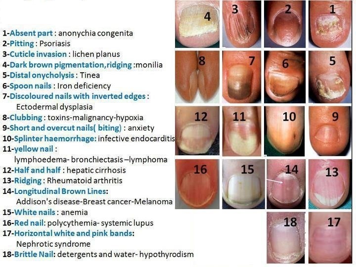
    33. Functions of skeleton system


    34. Skeletal Disorders
    • Osteoporosis-অস্টিওপোরোসিস
    • Osteomyelitis-অস্টিওমিলাইটিস
    • Osteopenia-অস্টিওপেনিয়া
    • Arthritis
    • Rickets/ Osteomalacia-রিকেট / অস্টিওমালাসিয়া
    • Gout-গাউট
    • Osteosarcoma-অস্টিওসারকোমা

    35. Classification of muscles

    Skeletal muscles are attached to bones by tendons, and they produce all the movements of body parts in relation to each other.

    Smooth muscle, found in the walls of the hollow internal organs such as blood  vessels, the gastrointestinal  tract , bladder , and uterus , is under control of the autonomic nervous system.

    Cardiac muscle is one of three types of vertebrate muscles, with the other two being skeletal and smooth muscles. It is involuntary, striated muscle that constitutes the main tissue of the walls of the heart.

    36. Functions of muscles
    • Movement
    • Maintenance of posture
    • Respiration
    • Heat generation
    • Pumping blood
    • Digestion
    • Urination
    • Childbirth
    • Vision
    37. What are the Muscular Disorders
    • Muscular dystrophy
    • Myalgia
    • Myopathy
    • Myositis
    • Muscle paralysis
    • Spasm or cramp

    38. What are the common body fluids

    Body fluids are liquids originating from inside the bodies of living humans. They include fluids that are excreted or secreted from the body.


    39. What are the fluid compositions

    40. Functions of body fluids


    41. What are the part of Immune system?
    The main parts of the immune system are:
    • White blood cells
    • Antibodies
    • Complement system
    • Lymphatic system
    • Spleen
    • Bone marrow
    • Thymus.
    41. What is Immunity? Classification of Immunity?
    Immunity is described as ability of the body to recognize, neutralize, or destroy harmful foreign substances in our body.

    42. What is antigens? What are the type of antigen?
    • Any substance when introduced into the body stimulate the production of antibody and react with the specific antibody and antigen receptors present on lymphocytes are called antigen.

    Classification of antigen
    Depending on origin three types-
    1. Exogenous antigens-entered the body from the outside, for example by inhalation, ingestion or injection (usually not virus)
    2. Endogenous antigens-generated within normal cells (viral or intracellular bacterial infection)
    3. Autoantigens (self protein or complex of proteins (and sometimes self DNA or RNA))
    43. What is antibody? What are the type of antibody?
    • Antibody is a large protein, constitiutes y-gloublin produced by plasma cells
    • It is used by the immune system to identify and nutralize pathogens such as bacteria and viruses
    • Antibodies are also celled immunogloublins
    • The antibody recognizes a unique molecule of the harmful agent called ANTIGEN, via the variable region

    44. What are the type of immune disorders?

    Three common autoimmune diseases are:
    • Type 1 diabetes. The immune system attacks the cells in the pancreas that make insulin.
    • Rheumatoid arthritis. This type of arthritis causes swelling and deformities of the joints.
    • Lupus. This disease that attacks body tissues, including the lungs, kidneys, and skin.
    45. What are the type of Immunodeficiency disorders?

    There are two types of immunodeficiency disorders:

    Primary: These disorders are usually present at birth and are genetic disorders that are usually hereditary. They typically become evident during infancy or childhood. However, some primary immunodeficiency disorders (such as common variable immunodeficiency) are not recognized until adulthood. There are more than 100 primary immunodeficiency disorders. All are relatively rare.

    Secondary: These disorders generally develop later in life and often result from use of certain drugs or from another disorder, such as diabetes or human immunodeficiency virus (HIV) infection. They are more common than primary immunodeficiency disorders.

    46. What is inflammation? Write down the 5 cardinal signs of inflammation.

    Inflammation is a process by which your body's white blood cells and the things they make protect you from infection from outside invaders, such as bacteria and viruses.

    There are five symptoms that may be signs of an acute inflammation:
    1. Redness
    2. Heat
    3. Swelling
    4. Pain
    5. Loss of function
    47. What is Phagocytosis? Write down of Phagocytosis stage.

    Phagocytosis, process by which certain living cells called phagocytes ingest or engulf other cells or particles. The phagocyte may be a free-living one-celled organism, such as an amoeba, or one of the body cells, such as a white blood cell.

    Stages of phagocytosis:
    1. Chemotaxis and adherence of microbeto phagocyte.
    2. Ingestion of microbe by phagocyte.
    3. Formation of phagosome.
    4. Fusion of the phagosome with a lysosome to form a phagolysosome.
    5. Digestion of ingested microbe by enzymes.
    6. Formation of residual body containing indigestible material.
    7. Discharge of waste materials. 

    48. What is autoimmune disease? Write down some autoimmune disease name.

    An autoimmune disease is a condition in which your immune system mistakenly attacks your body. The immune system normally guards against germs like bacteria and viruses. When it senses these foreign invaders, it sends out an army of fighter cells to attack them.

    1. Type 1 diabetes
    2. Rheumatoid arthritis (RA)
    3. Psoriasis/psoriatic arthritis
    4. Multiple sclerosis
    5. Systemic lupus erythematosus (SLE)
    6. Inflammatory bowel disease
    7. Addison’s disease
    8. Graves’ disease
    9. Sjogren’s syndrome
    10. Hashimoto’s thyroiditis
    11. Myasthenia gravis
    12. Autoimmune vasculitis
    13. Pernicious anemia
    14. Celiac disease
    49. Part of the lymphatic system.
    • Lymph
    • Lymph vessels
    • Lymph nodes
    • Bone marrow
    • Thymus
    • Spleen
    • Tonsils
    50. Function of the lymphatic system.
    • Lymph carries protein and large particulate matter away from the tissue space.
    • End products of digestion are absorbed mainly by lymph channels.
    • Important role in redistribution of fluid in the body.
    • Bacteria, toxins and other foreign bodies are removed from the tissues.
    • Maintenance of structural and functional integrity of tissue.
    • In immune response of the body.
    • Production and maturation of lymphocytes.
    51. Write down lymphatic disorders.
    • Lymphadenitis
    • Cancer
    • Anaphylactic shock
    • HIV/AIDS
    • Hodgkin's disease
    • Infectious mononucleosis
    • Lymphedema
    • Tonsillitis
    • Lupus erythematosus
    • Scleroderma
    52. What is Acid based balance?

    Acid-base balance refers to the mechanisms the body uses to keep its fluids close to neutral pH (that is, neither basic nor acidic) so that the body can function normally.

    53. What is Electrolyte balance?

    Electrolytes are minerals in the body that have an electric charge. They are in blood, urine and body fluids. Maintaining the right balance of electrolytes helps your body's blood chemistry, muscle action and other processes. Sodium, calcium, potassium, chlorine, phosphate and magnesium are all electrolytes.


    54. What is pH?
    • pH is a unit of measure which describes the degree of acidity or alkalinity (basic) of a solution.
    • It is measured on a scale of 0 to 14.
    • The formal definition of pH is the negative logarithm of the hydrogen ion activity.
    • pH = -log [H+]
    55. What are the causes of Electrolyte imbalance?

    The things that most commonly cause an electrolyte imbalance are:
    • vomiting
    • diarrhea
    • not drinking enough fluids
    • not eating enough
    • excessive sweating
    • certain medications, such as laxatives and diuretics
    • eating disorders
    • liver or kidney problems
    • cancer treatment
    • congestive heart failure
    56. Importance of Nutrition ?

    A healthy diet throughout life promotes healthy pregnancy outcomes, supports normal growth, development and ageing, helps to maintain a healthy body weight, and reduces the risk of chronic disease leading to overall health and well-being.

    57. What are the Nutritional deficiency disorder disease?


    Ads 728x90